solid mass on ovary
Imaging the suspected ovarian malncy: 14 cases TEN MDedge ObGynovarianmdedge.comType:PNGImages may be subject to copyright.

Adnexal Masses: US Characterization and Reporting | Radiology
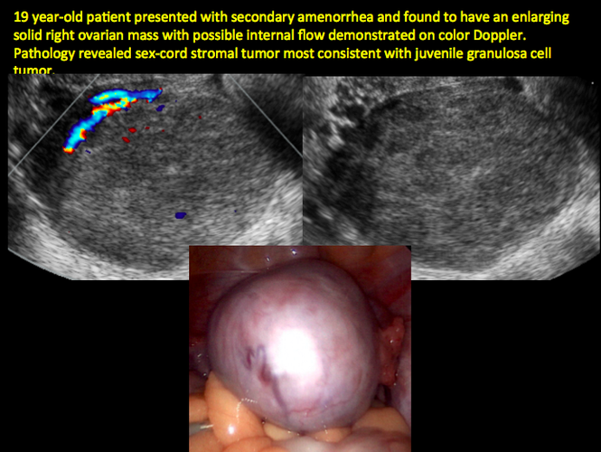
Imaging the suspected ovarian malignancy: 14 cases | MDedge ObGyn

Adnexal Masses: US Characterization and Reporting | Radiology
Ovarian Tumors What is the clinical setting when you will consider an ovarian mass? You would consider an ovarian mass, in any woman who comes in complaining of pressure symptoms. These symptoms include urinary frequency, pelvic discomfort, and ...

First International Consensus Report on Adnexal Masses: Management Recommendations - Glanc - 2017 - Journal of Ultrasound in Medicine - Wiley Online Library
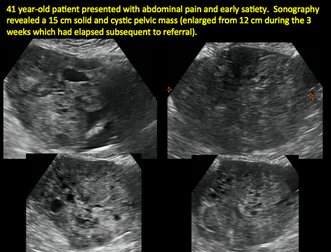
Imaging the suspected ovarian malignancy: 14 cases | MDedge ObGyn
Ultrasonography of adnexal masses: imaging findings

Simple vs. Complex Ovarian Cysts: The Link to Ovarian Cancer | Empowered Women's Health

Large right sided solid vascular ovarian mass in a 24-year-old... | Download Scientific Diagram
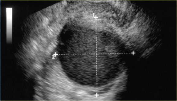
The Radiology Assistant : Common ovarian cystic lesions

Ovarian mass–differentiating benign from malignant: the value of the International Ovarian Tumor Analysis ultrasound rules - American Journal of Obstetrics & Gynecology

WK 5 L 1 Dysgerminoma The left ovary shows a solid mass with hypoechoic somewhat irregular septa dividing the tumor tissue into a lo… | Ultrasound, Ovarian, Ovaries
ovarian cysts, chocolate cysts

Gynaecology | 3.2 Adnexa : Case 3.2.4 Malignant ovarian lesions | Ultrasound Cases
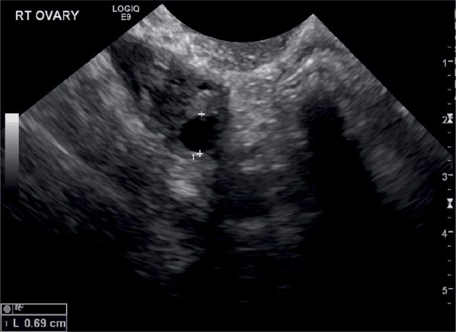
Sonographic Assessment of Ovarian Cysts and Masses (Chapter 8) - Gynaecological Ultrasound Scanning

Adnexal Masses: US Characterization and Reporting | Radiology

Between the uterus and the right ovary, 5.57×2.91 cm sized solid mass... | Download Scientific Diagram
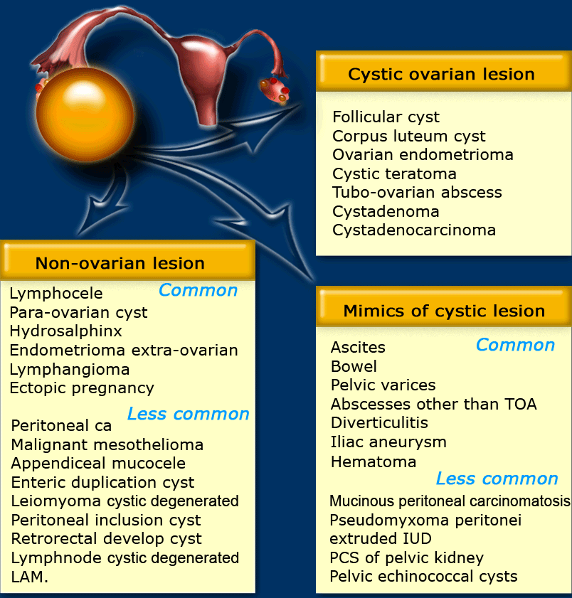
The Radiology Assistant : Roadmap to evaluate ovarian cysts
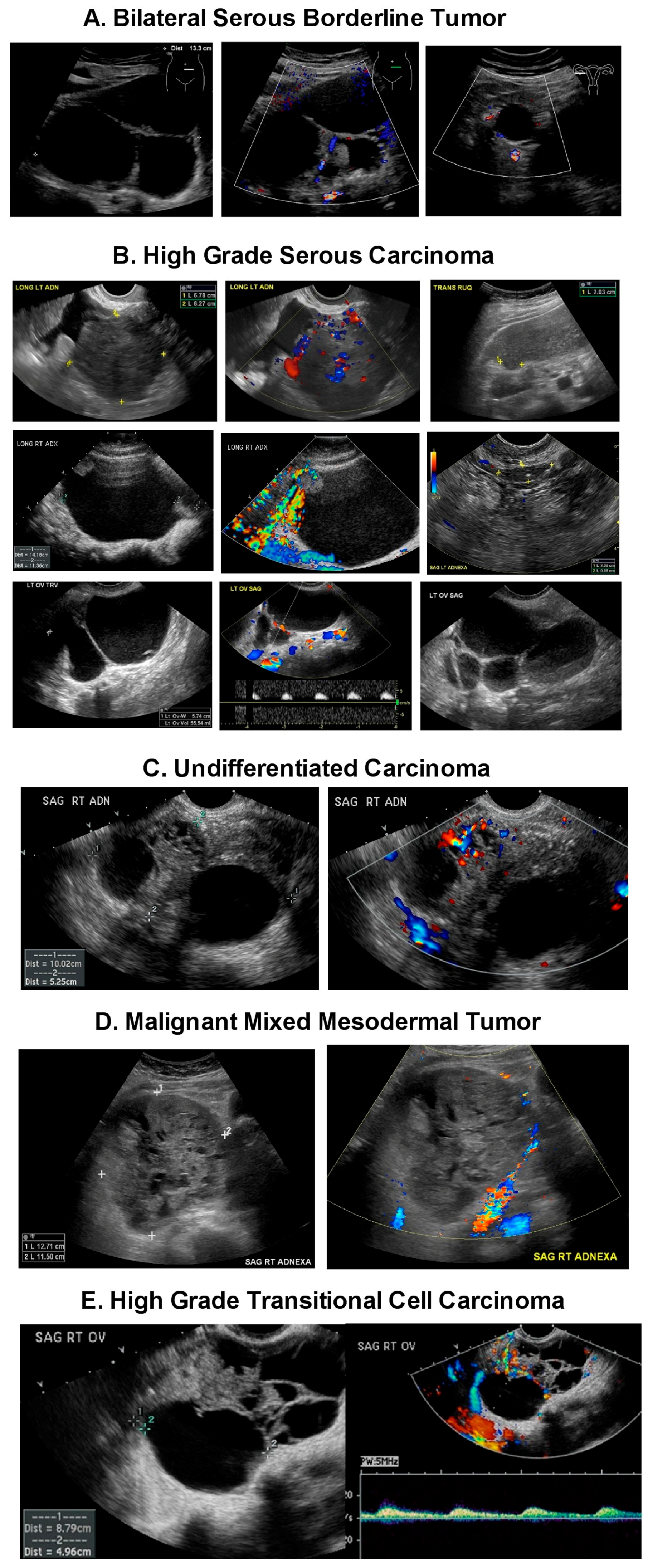
Diagnostics | Free Full-Text | Ultrasound Monitoring of Extant Adnexal Masses in the Era of Type 1 and Type 2 Ovarian Cancers: Lessons Learned From Ovarian Cancer Screening Trials | HTML
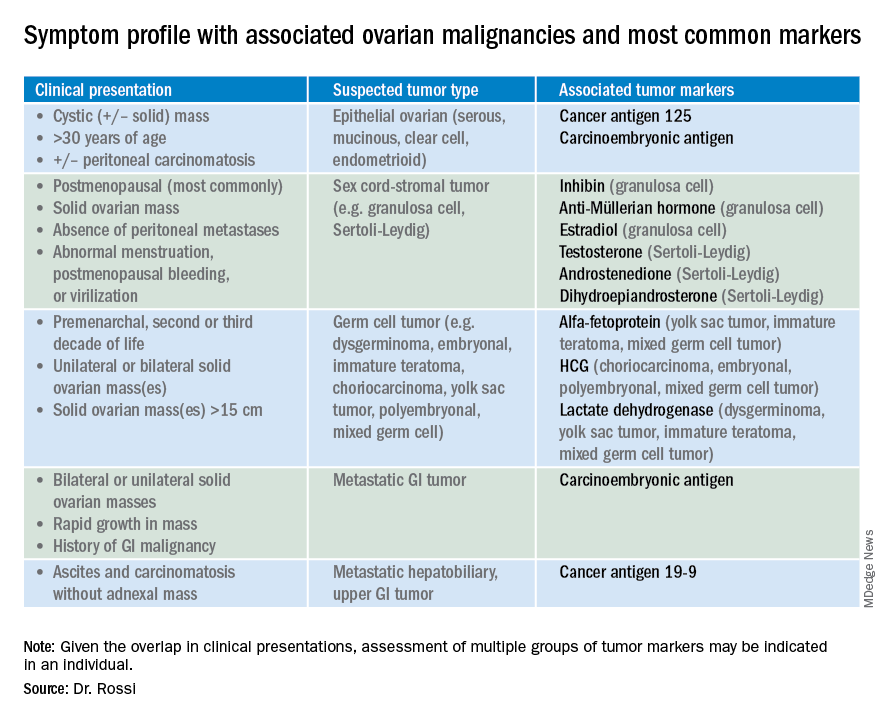
Ovarian tumor markers: What to draw and when | MDedge ObGyn

Adnexal Masses: US Characterization and Reporting | Radiology
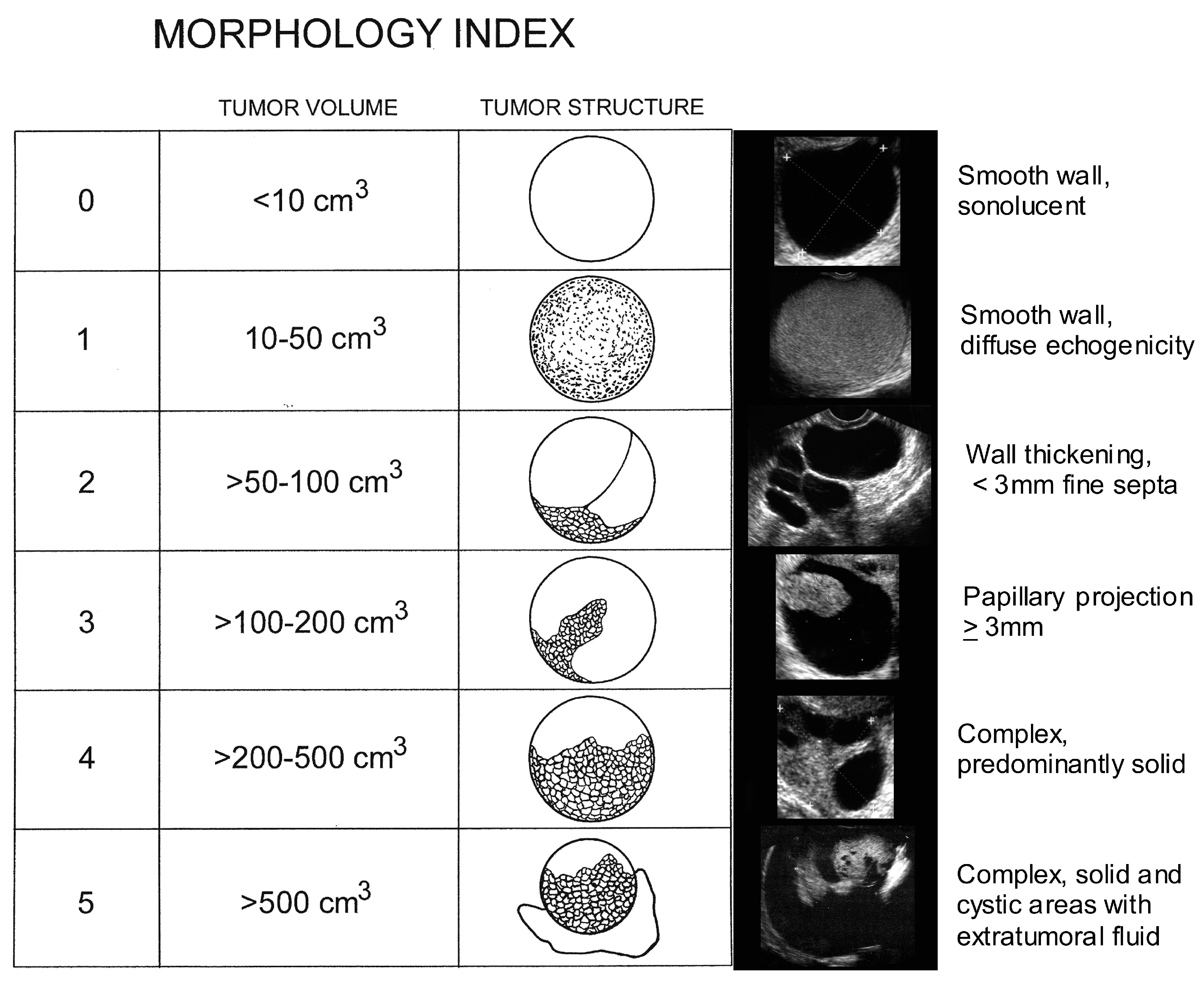
Diagnostics | Free Full-Text | Ultrasound Monitoring of Extant Adnexal Masses in the Era of Type 1 and Type 2 Ovarian Cancers: Lessons Learned From Ovarian Cancer Screening Trials | HTML

Ultrasonography
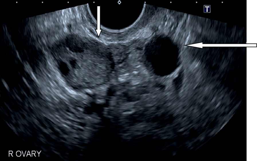
Sonographic Assessment of Ovarian Cysts and Masses (Chapter 8) - Gynaecological Ultrasound Scanning
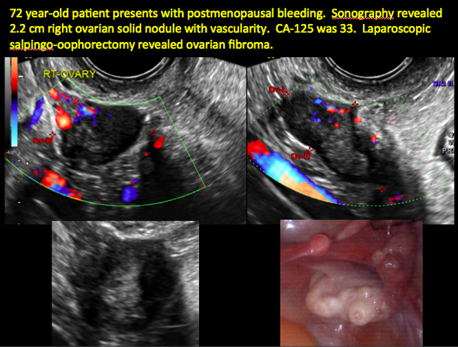
Imaging the suspected ovarian malignancy: 14 cases | MDedge ObGyn
A Case of a Giant Ovarian Mass in a Teenager: Case Report and Review of Literature

Transabdominal ultrasound of right ovarian mass. a Right ovarian mixed... | Download Scientific Diagram
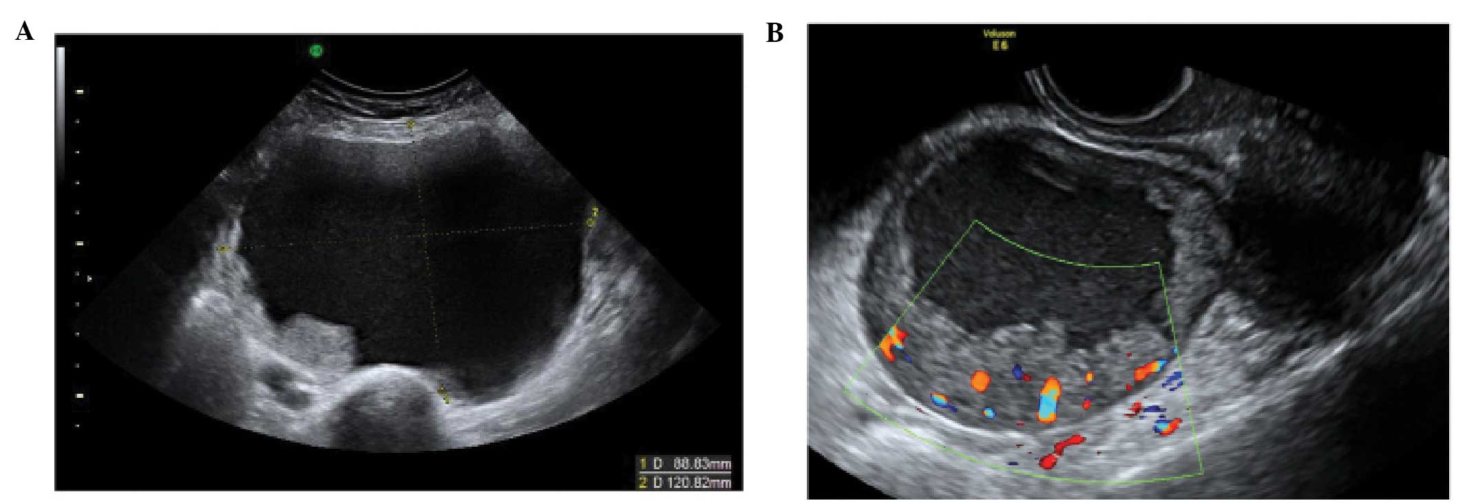
The characteristic ultrasound features of specific types of ovarian pathology (Review)

Ovarian Cysts & Tumors Fort Worth - FW Center for Pelvic Medicine
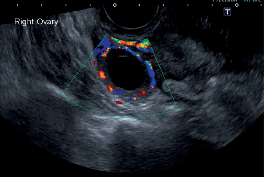
Sonographic Assessment of Ovarian Cysts and Masses (Chapter 8) - Gynaecological Ultrasound Scanning
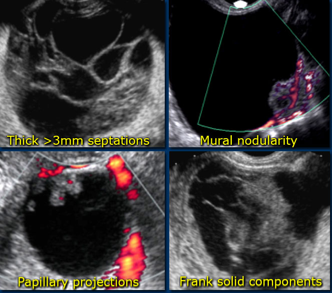
The Radiology Assistant : Roadmap to evaluate ovarian cysts

Gynaecology | 3.2 Adnexa : Case 3.2.4 Malignant ovarian lesions | Ultrasound Cases
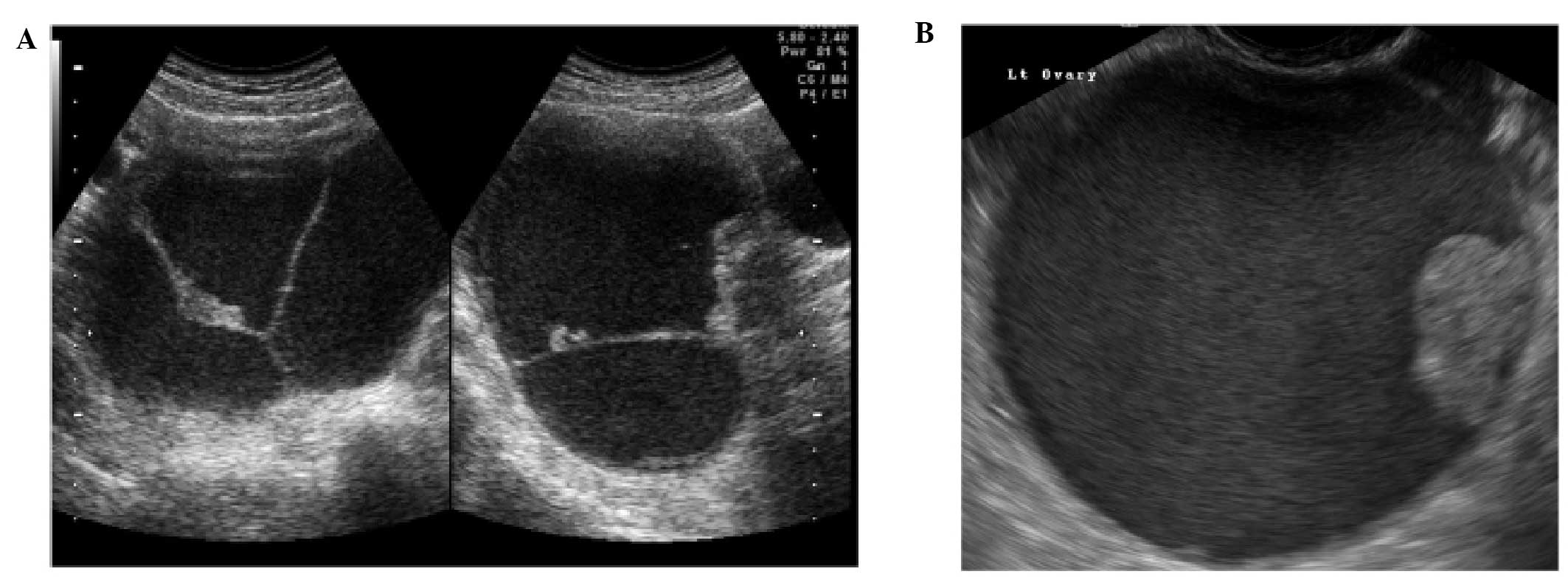
The characteristic ultrasound features of specific types of ovarian pathology (Review)

EPOS™ - C-0354

Simple vs. Complex Ovarian Cysts: The Link to Ovarian Cancer | Empowered Women's Health

Ovarian Cysts
Ovarian Leydig cell tumor in a post-menopausal patient with severe hyperandrogenism

Ovarian Cysts

3.2.4 Malignant ovarian lesions | Ultrasound Cases

Imaging Evaluation of Ovarian Masses | RadioGraphics
Posting Komentar untuk "solid mass on ovary"